When I first began analyzing granulation tissue wound pictures during direct wound care, I quickly realized these images provide much more than surface-level information. They act as visual markers of recovery, helping caregivers and patients recognize whether healing is progressing smoothly or if complications may be emerging.
From my own experience, wounds showing moist, beefy-red tissue usually signal strong healing with healthy blood vessel growth. By contrast, pale, gray, or friable tissue can point to circulation issues, infection risk, or stalled recovery. Recognizing these subtle differences early can prevent setbacks and promote faster outcomes.
This article explains how wound pictures provide visual insights into the healing process, offers guidance for interpretation, and shares real-world examples to help patients and caregivers build confidence in wound management.
Top Takeaways
- Wound photos are healing checkpoints—they track stages and flag concerns.
- Healthy vs. unhealthy tissue is visible—red and moist = progress; pale or uneven = warning.
- Research confirms benefits—photos improve outcomes and keep patients engaged.
- Photos build trust—they show progress clearly to both patients and caregivers.
How Wound Pictures Show Healing
Granulation tissue wound pictures act like a timeline of the body’s repair process.
- Healthy signs: Red or pink, moist, slightly bumpy tissue with active blood supply.
- Warning signs: Pale, grayish, dry, or irregular tissue—often linked to poor circulation or infection.
By reviewing images regularly, caregivers can evaluate whether the wound is shrinking, stable, or worsening, and adjust treatment strategies accordingly, while also watching for warning signs such as purulent drainage, which may indicate infection and require immediate medical attention.
Expert Insight
"In my wound care practice, I’ve found that photos are more than documentation—they’re progress reports. A healthy red tissue bed signals steady rebuilding, while pale or uneven tissue is an early red flag. These visual insights often guide critical decisions that written notes might miss."
Case Studies & Real-World Examples
Post-Surgical Healing
Patient: 54-year-old after abdominal surgery
Early signs: Pale, uneven tissue suggesting poor circulation
Interventions: High-protein diet, improved dressings, mobility exercises
Outcome: By week 4, photos showed strong red granulation tissue
Takeaway: Images provided reassurance and proof of progress for both patient and provider
Diabetic Foot Ulcer
Patient: Long-standing diabetic ulcer
Challenge: Unhealthy, friable tissue visible in weekly photos
Interventions: Debridement and infection management
Outcome: Transitioned to healthy red tissue in several weeks
Takeaway: Regular photo documentation guided timely care and motivated the patient
Research Perspective
Studies confirm: Documenting wounds with photos improves decision-making and outcomes
Clinical experience: Photos serve as educational tools that empower patients to recognize healing vs. warning signs
Patient: 54-year-old after abdominal surgery
Early signs: Pale, uneven tissue suggesting poor circulation
Interventions: High-protein diet, improved dressings, mobility exercises
Outcome: By week 4, photos showed strong red granulation tissue
Takeaway: Images provided reassurance and proof of progress for both patient and provider
Patient: Long-standing diabetic ulcer
Challenge: Unhealthy, friable tissue visible in weekly photos
Interventions: Debridement and infection management
Outcome: Transitioned to healthy red tissue in several weeks
Takeaway: Regular photo documentation guided timely care and motivated the patient
Studies confirm: Documenting wounds with photos improves decision-making and outcomes
Clinical experience: Photos serve as educational tools that empower patients to recognize healing vs. warning signs
Supporting Statistics
Chronic wounds are widespread
8.2 million Medicare beneficiaries experience wounds annually, costing $28–$96 billion
Source: NIH – Chronic Wounds Study
Pressure ulcers are common
More than 2.5 million people in the U.S. develop them annually
Source: AHRQ – Pressure Ulcer Facts
Diabetic foot ulcers are high-risk
12–25% of people with diabetes will develop a foot ulcer in their lifetime
Source: NCBI – Diabetic Foot Ulcers
Long-term risks are severe
Up to 65% recurrence within 5 years, with amputation and mortality risks as high as 70%
Source: American Diabetes Association – Foot Complications
Photography is clinically supported
Recognized as a valuable complement to written wound documentation
Source: PubMed – Wound Photography Guidelines
Final Thought & Opinion
Granulation tissue wound pictures are much more than records—they are active tools that show the story of healing in real time. They make recovery visible, help patients feel encouraged, and allow caregivers to act quickly when issues arise.
From my perspective, photos often reveal what words cannot. They highlight subtle improvements, motivate patients to stay consistent, and bring clarity to situations where uncertainty is common. In short, wound pictures are essential for effective healing, trust, and better outcomes—especially when paired with the right wound care products that support each stage of recovery.
Next Steps
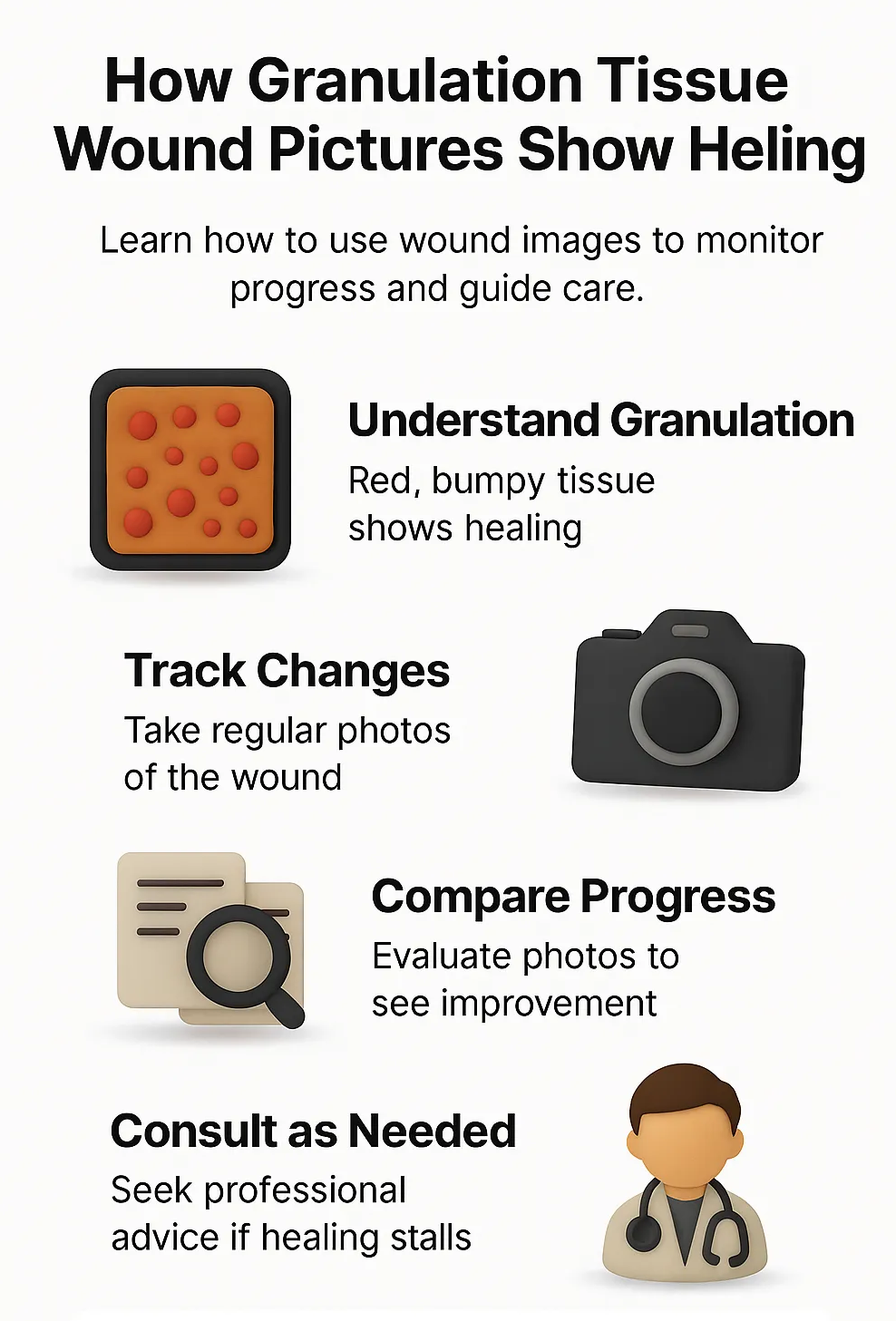
Compare your wound: Healthy = moist, red tissue; warning = pale, gray, or uneven tissue
Track progress: Take consistent photos (same lighting, angle, and timing)
Share with your provider: Use images to guide professional care decisions
Stay informed: Rely on credible sources like NIH, CDC, and ADA
Act quickly: Contact a wound care specialist if photos reveal infection or stalled healing